Home
/ Muscles In The Body Diagram : Skeletal Muscle Wikipedia / This is what happens in the body.
Muscles In The Body Diagram : Skeletal Muscle Wikipedia / This is what happens in the body.
Muscles In The Body Diagram : Skeletal Muscle Wikipedia / This is what happens in the body.. Diagrams of the muscles and guide to how they work. These muscles are the only voluntary muscles in the body—we can control these muscles. There are some 700 named muscles in the body, and other smaller muscle tissues that are part of. Muscles, connected to bones or internal organs and blood vessels, are in charge for movement. Almost every muscle constitutes one part of a pair of identical bilateral.
Diagrams of the muscles and guide to how they work. Muscle charts of the huma. These include mobility, stability, posture, circulation, digestion, and more. Here are five other facts to keep in mind about the muscular system. The muscles of the human body are responsible for movement;
Amazon Com Laminated 24x24 Poster Anatomy Of Human Body Parts Body Parts Names Human Anatomy Human Anatomy Diagram Human Anatomy Everything Else from images-na.ssl-images-amazon.com Human body muscle system, the muscles of the human body that work the skeletal system, that are under voluntary control, and that are concerned with movement, posture, and balance. Muscle diagram male body names. The sartorius muscle is positioned more superficially than the other in the leg muscles. These include mobility, stability, posture, circulation, digestion, and more. They maintain posture and provide the strength for lifting and pushing. These muscles are able to move the upper limb as they originate at the vertebral column and insert onto. Anterior muscles in the body. Part of quadriceps group, extends leg at knee.
These muscles hold the inner ear together and are connected to.
There are some 700 named muscles in the body, and other smaller muscle tissues that are part of. This muscle diagram is interactive: Just a little deeper of biceps brachii lies brachialis muscle that helps in flexing the elbow. Labeled vector illustration chart on white background. Almost every movement in the body is the outcome of muscle contraction. Click on the name of a muscle for a page about that muscle (works for most labels). Within this group of back muscles you will find the latissimus dorsi, the trapezius, levator scapulae and the rhomboids. There are around 650 skeletal muscles within the typical human body. But, your soleus muscle in your lower leg and muscles in your back involved in maintaining posture contain mainly slow twitch muscle fibres. Each of these muscles is a discrete organ constructed of skeletal muscle tissue blood vessels tendons and nerves. Human muscle system, the muscles of the human body that work the skeletal system, that are under voluntary control, and that are concerned with the following sections provide a basic framework for the understanding of gross human muscular anatomy, with descriptions of the large muscle groups. It does work independently but it actually supports biceps brachii to flex the elbow joint. Diagrams of the muscles and guide to how they work.
Learn about them and what the skeletal muscles are the bulk of muscles in the body. It also helps raise the body from a supine. Within this group of back muscles you will find the latissimus dorsi, the trapezius, levator scapulae and the rhomboids. Each of these muscles is a discrete organ constructed of skeletal muscle tissue blood vessels tendons and nerves. Muscle charts of the huma.
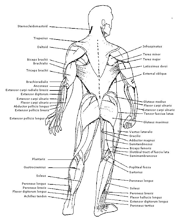
Physiology Identification Of Muscles On The Human Body from www.brianmac.co.uk Below are two human body muscle diagrams, showing the front and back of the body. Within this group of back muscles you will find the latissimus dorsi, the trapezius, levator scapulae and the rhomboids. Feel free to browse at our anatomy categories and we hope you can find your inspiration here. Muscles, connected to bones or internal organs and blood vessels, are in charge for movement. Thank you for visiting major muscles of the body diagram pictures. Heaviest muscle in body, extends/straightens leg at hip during walking. Despite their similar names, teres major has different actions and innervation from the teres minor. This is a table of skeletal muscles of the human anatomy.
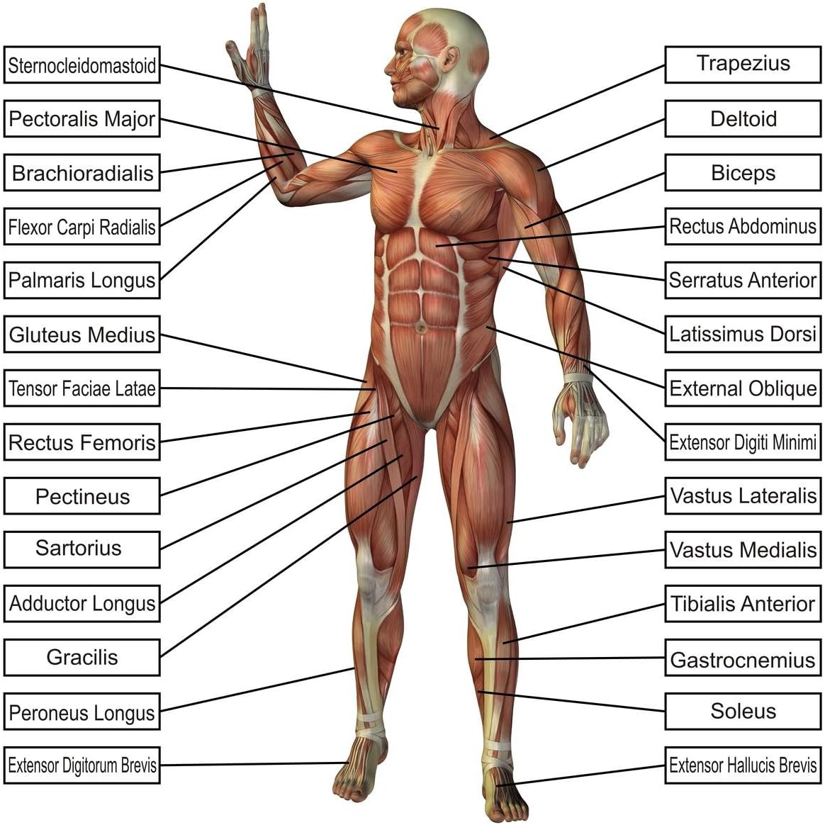
The muscles labelled in the anterior muscles diagram shown above are listed in bold in the following table
The teacher should show the students the muscle cards and explain. These muscles are in fact a bundle of muscles that share a common insertion point near the elbow joint. If you found any images copyrighted to yours, please contact us and we. Just a little deeper of biceps brachii lies brachialis muscle that helps in flexing the elbow. Teres major is a thick and ovoid muscle in the upper arm. This muscle diagram is interactive: The next life study seated female figure, shows the upper part of the pectoralis major positioned flat against the rib cage, with very the muscle helps bend the torso forward in the movement known as the flexion of the vertebral column. They maintain posture and provide the strength for lifting and pushing. The human muscular system is an organ system composed of skeletal muscles, smooth muscles, and cardiac muscles. Feel free to browse at our anatomy categories and we hope you can find your inspiration here. But, your soleus muscle in your lower leg and muscles in your back involved in maintaining posture contain mainly slow twitch muscle fibres. There are approximately 640 skeletal muscles within the typical human, and almost every muscle constitutes one part of a pair of identical bilateral muscles, found on both sides, resulting in approximately 320 pairs of muscles, as presented in this article. The ear contains the smallest muscles in the body alongside the smallest bones.
This is a table of skeletal muscles of the human anatomy. Located immediately below the skin) muscles of the body. Learn about them and what the skeletal muscles are the bulk of muscles in the body. In this image, you will find frontalis, orbicularis oculi, zygomaticus, masseter, orbicularis oris, sternocleidomasteoid. It does work independently but it actually supports biceps brachii to flex the elbow joint.
Anatomy Muscles Man In Body Vtwctr from i0.wp.com It permits movement of the body, maintains posture and circulates blood throughout the body. The ear contains the smallest muscles in the body alongside the smallest bones. These muscles are able to move the upper limb as they originate at the vertebral column and insert onto. The sartorius muscle is positioned more superficially than the other in the leg muscles. Within this group of back muscles you will find the latissimus dorsi, the trapezius, levator scapulae and the rhomboids. Each of these muscles is a discrete organ constructed of skeletal muscle tissue blood vessels tendons and nerves. Published december 28, 2017 at 768 × 1024 in so…what can you feel or move? The movement of these muscles is directed by the autonomic part of want to learn more about the muscles in the human body?
This is what happens in the body.
See how all sharpness disappears? Just a little deeper of biceps brachii lies brachialis muscle that helps in flexing the elbow. This is what happens in the body. There are around 650 skeletal muscles within the typical human body. Muscle charts of the huma. Heaviest muscle in body, extends/straightens leg at hip during walking. Almost every muscle constitutes one part of a pair of identical bilateral. It permits movement of the body, maintains posture and circulates blood throughout the body. The superficial back muscles are the muscles found just under the skin. This is a table of skeletal muscles of the human anatomy. Within this group of back muscles you will find the latissimus dorsi, the trapezius, levator scapulae and the rhomboids. To get started, choose a muscle group either on the muscle chart or in the muscle list on this page. On the board, the teacher should place a picture of the diagram of the human body.